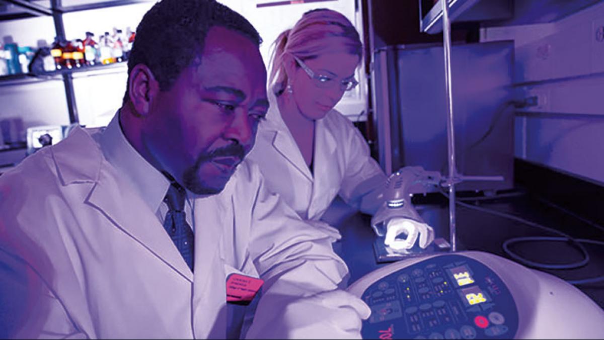
What if there was a way to treat debilitating diseases without drugs or surgery? What if chronic injuries could be healed with the application of something as ubiquitous as light?
Scientists have known for years that some wavelengths of light in certain doses can heal tissue, but they are only now uncovering exactly how light accomplishes its therapeutic effects.
Known as phototherapy, the use of light in medical treatment is producing surprisingly successful results in the treatment of a variety of ailments from topical infections and chronic wounds to autoimmune and chronic degenerative diseases, says Chukuka S. Enwemeka, dean of the University of Wisconsin–Milwaukee’s College of Health Sciences. Enwemeka, who is internationally known for his work in phototherapy, is one in a cluster of scientists at UW–Milwaukee (UWM) conducting studies in this emerging field of medicine.
Work by the UWM researchers focuses on wavelengths of light that lie in two regions of the electromagnetic spectrum: longer wavelengths in the far-red to near-infrared (NIR) region and shorter wavelengths in the visible blue region of the spectrum.
VISIBLE SPECTRUM
Light is energy, a kind of radiation consisting of photons whose individual energy levels correspond to specific wavelengths. On the electromagnetic spectrum, light is arranged by wavelength along with other forms of energy, including gamma rays, microwaves, and radio waves. White light, or the “visible spectrum,” exemplified by the sun or incandescent light bulbs, includes blue, yellow and red light and makes up only a tiny section of the electromagnetic spectrum. White light lies between ultraviolet radiation and near red and infrared radiation.
 Determining the best wavelength for phototherapy is a difficult task, says Jeri-Anne Lyons, a UWM associate professor of biomedical sciences. “[We use] only certain wavelengths, at a certain intensity, for a certain amount of time.” Studies show that though red to near-infrared light covers wavelengths of about 600 to 1100 nanometers (nm), the 670 nm and 830 nm wavelengths are the most beneficial of the near-infrared (NIR) spectrum. Because light in these wavelengths can penetrate the skin and be absorbed by subcutaneous cells, it can act on wounds, internal injuries, and disease.
NIR light—which produces no heat—is administered through an array of light-emitting diodes (LEDs) that can be configured to the desired wavelength. Sometimes LEDs are arranged in large, flat arrays to facilitate the treatment of large wounds.
Finding the appropriate dose and dose regimen for delivering the light is important. “Like ingested medication, it’s all about the dose,” says Lyons, adding that establishing dosage for near-infrared light has been largely a matter of trial and error.
“We started irradiating damaged cells in cultures and found what appeared to be a ‘sweet spot’ in terms of dosage,” says Janis Eells, UWM professor of biomedical sciences who studies how NIR light helps to slow degenerative eye disease. This dosage, which stimulated repair in the cultured cells, was also shown to be effective in animal models of disease. “We are conducting dose-response experiments now to determine the optimal dose of light.”
Last year, Eells and a team of collaborators conducted a wound-healing study in spinal cord injured veterans who, because of being bedridden and inactive, developed pressure ulcers, or bedsores, that wouldn’t heal. “Chronic wounds are ‘stuck’ in the inflammatory phase of healing. NIR light removes that obstacle,” says Eells. “If you can tone down the inflammation in a non-healing wound, like a pressure ulcer, you speed the healing.”
Conducted at the Zablocki Veteran Affairs Medical Center in Milwaukee, the study compared the rate of wound healing in two groups of veterans with similar ulcers. All wounds were first treated for four weeks with standard care (keeping the wound clean and free of infection). But only one group was subsequently given phototherapy three times a week for ninety seconds for four weeks. According to the study, the rate of healing was 250% faster in the wounds receiving the NIR light.
CELLULAR RECEPTION
Although study at the Zablocki Medical Center showed that phototherapy holds tremendous promise, there is much to be done before the treatment becomes standard practice in America. This includes phototherapy experiments that help researchers better understand the cellular processes involved.
For any light therapy to work, there must be light-responsive molecules within the body that are positively altered in some way by the light waves’ energy. UWM researchers have built upon the work of Tiina Karu at the Russian Academy of Laser Sciences. It was Karu who determined that far-red and NIR light, applied using low-intensity lasers, acts on organelles of human cells known as mitochondria. More specifically, the light acts upon a molecule called cytochrome c oxidase.
A mammalian cell contains thousands of mitochondria, protein “machines” in charge of converting the energy in the food we eat into a form of chemical energy that the cell can use. Research by Karu and others has shown that when cytochrome c oxidase, an enzyme that is part of the energy-generating sequence in the mitochondria, is excited by photons of NIR light, a number of cellular changes can occur. For example, there is an increase in the messages exchanged between mitochondria and the cell’s nucleus resulting in a boost in the mitochondria’s output of ATP, a molecule that increases the cell’s energy. This triggers release of signal molecules that tell genes to go into action. The genes activate release of antioxidants and other cell-protecting factors that counteract cell degeneration by repairing mitochondria that have become damaged or dysfunctional. Antioxidants also work to clean up free radicals, highly chemically reactive molecules that can bond to and alter other molecules in destructive ways related to aging and cancer.
“Through this enzyme [cytochrome c oxidase], the light gives a ‘molecular kick’ to the mitochondria, telling the cell to turn on a large number of antioxidant and energy-boosting genes,” summarizes Eells. She says that it is a concept that makes perfect sense, when you consider that “mitochondria are similar to chloroplasts in plants, which absorb red and blue wavelengths of light and use the light energy to make chemical energy [during] photosynthesis.”
 Eells, who has great admiration for the complex ways in which mitochondria behave and communicate with the rest of the cell, says that these organelles do far more than simply produce energy for cells. “They not only control the life of the cell, they control cell death too,” she says. “If a cell becomes diseased or dysfunctional, the mitochondria send out signals which tell the cell to self-destruct in an organized fashion—so that it doesn’t take out its neighbors at the same time.”
When mitochondrial function is disrupted, says Eells, unintentional cell death occurs. Additionally, the stage is set for another imbalance: over-production of free radicals. In limited numbers, free radicals play important roles in cell communication. However, these molecules have a dark side. An accumulation of too many creates what’s called “oxidative stress” and will damage other components of the cell, such as proteins and DNA.
Near-infrared light can help restore balance by activating antioxidant molecules that “disarm” the free radicals and help repair and even reverse their damage. This restorative component of NIR therapy has interesting implications for slowing disease impacts and even some aspects of aging.
Eells has seen this kind of restoration in her own work treating eye diseases like retinitis pigmentosa and diabetic retinopathy. She has found that NIR light triggers positive cellular responses that actually preserve many of the eye’s photoreceptor cells that these diseases would otherwise have destroyed.
ANTIBIOTIC EFFECTS
In contrast to far-red and NIR light which stimulate the body’s repair of injured cells, blue light improves wound healing by killing the bacteria that cause wound infections.
UWM’s Enwemeka is a pioneer in the use of LED blue light to clear infections. In a 2007 study supported by Dynatronics Corporation, Enwemeka discovered that some wavelengths of blue light, especially those in the 405–470 nm wavelength, kill bacteria so effectively that the process even works on MRSA, the antibiotic-resistant “superbug” form of Staphylococcus aureus.
What gives this shorter wavelength of light such a powerful antibiotic effect?
One explanation is that bacteria contain light-sensitive, iron-rich molecules that, upon absorbing blue light, generate free radicals that kill the bacteria. Enwemeka suggests that blue light also acts on cytochrome c oxidase in the cells’ mitochondria—like NIR light—but in this case causes the cytochrome to pair up with nitric oxide, creating a toxic environment for bacteria.
Blue light therapy has achieved undeniable laboratory results in treating antibiotic-resistant MRSA. Enwemeka demonstrated that one dose of blue light irradiation killed as much as 92% of two pervasive MRSA strains. He has also seen what he describes as astonishing results at a clinic he works with in Brazil where the blue light, combined with NIR light, is used to treat chronic wounds like diabetic ulcers.
Enwemeka notes that to achieve complete destruction of bacterial colonies with blue light, the treatment must be repeated. But given its relative ease of application, blue light therapy holds tremendous promise as an alternative to full-spectrum antibiotics, many of which are decreasing in efficacy. Humans are exposed to so many antibiotics—through food sources and careless use of prescribed medications—that bacterial resistance is now rising much faster than the rate of new drug discovery.
Unlike NIR light, which can penetrate below the skin, the shorter wavelength of blue light can only be absorbed at the surface of a wound. “But suppose that the [tissue] layer is thick,” says Enwemeka. “In that case, you have to increase the dosage [to reach all the bacteria]. Now the question is, ‘will using a higher dosage kill more normal cells along with the bacteria?’ ”
Enwemeka’s current project with UWM physicist Valerica Raicu aims to render that question moot. The two are developing new technology that will allow blue light’s wavelength to penetrate skin—and dense bacterial colonies—more deeply, the way NIR light can, but without increasing dosage. The goal is to achieve complete bacterial eradication in a wound.
Why doesn’t the blue light also kill the cells of the human host cells? While Enwemeka has found evidence that small numbers of the human cells in proximity to the infection are also killed after blue light irradiation, the answer is not yet clear. But it may have to do with differences in the photosensitive molecules present in bacteria versus mammalian cells.
In recent research, Enwemeka found that in higher doses, treatment with blue light yielded a completely unexpected result: While some human cells are destroyed by irradiation along with much of the bacteria, a greater percentage of human cells appear to multiply.
How can blue light both destroy and generate cells simultaneously?
Enwemeka doesn’t yet know, but the possibility isn’t as far-fetched as it may sound. His team is exploring the fact that one of the downstream effects of blue light therapy could be the activation of genes responsible for division of mammalian cells. Their research with cells in culture has shown that blue light exposure increases growth factors, which leads to proliferation of fibroblasts, a type of connective tissue cell that plays a role in wound healing.
Low-level light therapy has also been implicated in activating genes involved in programmed cell death and in the stress response generated by too many free radicals. Enwemeka’s team hypothesizes that both cell proliferation and cell death can occur together if there is more response in the genes controlling cell death than in those that stimulate growth factors.
BEYOND TRIALS
With so much success, why isn’t phototherapy being used more widely?
Lyons, for instance, has tested phototherapy as a method of controlling the severity of multiple sclerosis, an autoimmune disease that causes progressive paralysis—interrupting vision, balance, and even thought processes by destruction of nerve cells and the spinal cord.
But while the U.S. Food and Drug Administration has approved NIR light for the treatment of pain and depression as well as skin conditions like acne and psoriasis, the FDA has not yet approved it for wound healing or the treatment of diseases like multiple sclerosis.
“It’s considered ‘alternative’ therapy in Western medicine. It seems too simple for people to accept,” says Lyons.
Enwemeka agrees, but says that UWM research is moving phototherapy closer to mainstream acceptance. What the FDA is waiting for, he says, is confirmation from a large-scale clinical study before approving phototherapy for a wider variety of ailments. It’s something Enwemeka and his colleagues at UWM are determined to accomplish.


